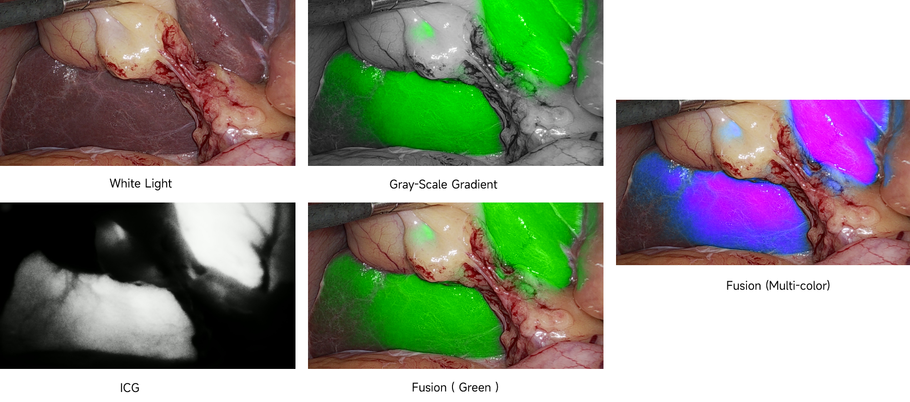
Near-infrared fluorescence imaging is a technology that uses near-infrared light to excite and emit fluorescence signals to achieve imaging. Through this technology, we can observe fine structures and biomolecule information that cannot be seen in the while light mode. Let’s take a closer look at the principles and applications of near-infrared fluorescence imaging!

Different image modes of 2nd Generation of Hikimaging MIK5 Fluorescence Solution
1.Principles of near-infrared fluorescence imaging
The main absorption peaks of hemoglobin and water are in the visible light region and the infrared region, and there is a light absorption trough in the near-infrared 650-900nm range [1], yet human tissue would absorb and radiate near-infrared light weaker than visible light. Therefore, by choosing light in the near-infrared 650-900nm range as excitation light and fluorescence, deep tissue (1-2cm) images can be obtained.

[1] R. A Weissleder, Nat. Biotechnol., 2001, 19, 316-317.
ICG(Indocyanine Green,)
ICG is one kind of near-infrared fluorescent dye approved by FDA for in vivo use. Among all NIR fluorescent probe types, ICG is the only clinical diagnostic NIR fluorescent material approved for liver function and ophthalmic angiography.
Advantage
- Both excitation light and fluorescence are in the near-infrared region and are within the optical window of the tissue, allowing NIR fluorescence imaging.
- Dry ICG is stable and easy to store
- Low toxicity, rapid excretion, effective binding to blood lipoproteins, will not leak from the blood circulation, and is safer for patients
- Easy to prepare, mass-produced, low cost
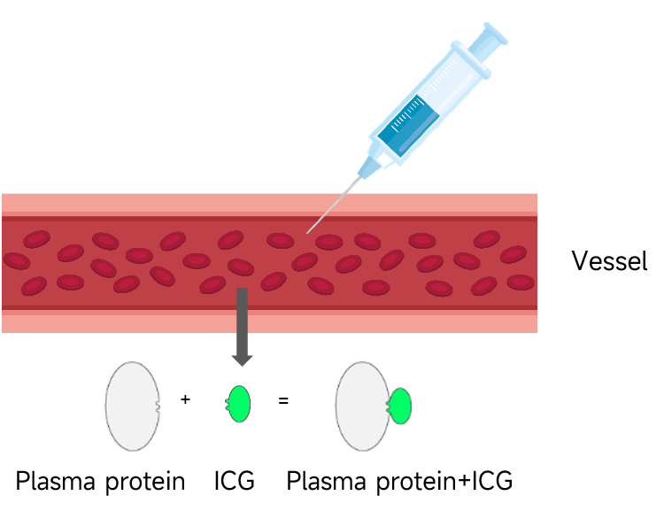
Adsorption phenomenon of ICG fluorescent molecules
The amphiphilic nature of ICG lipid allows it to enter the human body through intravenous injection and other methods, and can be free or combined with plasma proteins and transported throughout the body through blood and tissue fluid.

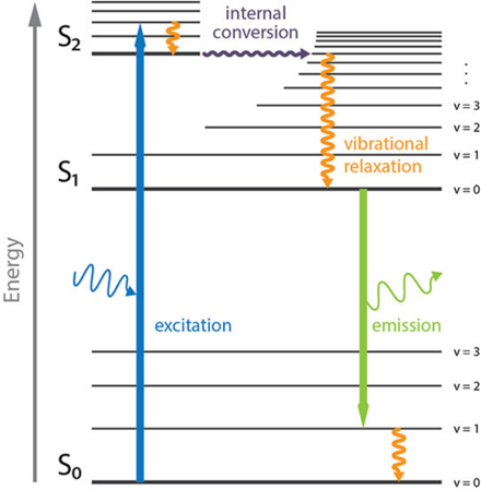
Indocyanine Green and its fluorescent reaction
Fluorescent molecules located in the lowest energy state (ground state), when irradiated by 700-800nm excitation light, absorb photon energy and are excited to transition to the excited state (absorption process).
Within the fluorescence lifetime, fluorescent molecules transition downward from the excited state to the ground state (emission process) with a certain probability, and emit fluorescence in the 820-900nm range.

The fluorescence characteristics are shown in the figure below. The light curve is the excitation light absorption response curve, and the dark curve is the fluorescence emission intensity curve. However, in actual scenarios, the fluorescence intensity will be significantly lower than the excitation light intensity.

2.The impact of ICG concentration
Lots of researches have been done to illustrate that the fluorescence intensity produced by ICG is related to various factors such as ICG concentration and solvent.
- Effect of concentration: If the concentration of ICG is too high, the ICG molecules in the solution will clump together to produce polymer ICG (the aggregation phenomenon of ICG fluorescent molecules). The fluorescence yield of polymer ICG is extremely low. At the same time, the presence of polymer ICG in the solution will cause free ICG molecules to form. The probability of receiving excitation light is reduced.
- Solvent influence: Different solvents have different ICG concentrations at which the fluorescence intensity reaches the maximum value.
- Therefore, higher concentrations are not always better, It is recommended to choose the appropriate concentration and injection method according to the clinical scenario.
Appendex: Introduction of ICG(Indocyanine Green)
ICG is one kind of near-infrared fluorescent dye approved by FDA for in vivo use. Among all NIR fluorescent probe types, ICG is the only clinical diagnostic NIR fluorescent material approved for liver function and ophthalmic angiography.
Physical properties:
1.Appearance: dark green cyan or dark brown red powder;
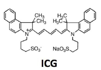
2.Polycyclic structure, and a sulfate group is connected to each polycyclic structure. The polycyclic structure has strong lipophilicity, while sulfuric acid, solubility: soluble in water, DMSO, methanol, ethanol (slightly soluble);
3.Lipid-water amphiphilicity: The two ends of a carbon bond in the ICG molecule are connected to two groups and have a certain degree of water solubility. This complex molecular structure ultimately leads to the ICG molecule being amphipathic, that is, it is both lipophilic and hydrophilic.
Advantage
- Both excitation light and fluorescence are in the near-infrared region and are within the optical window of the tissue, allowing NIR fluorescence imaging.
- Dry ICG is stable and easy to store
- Low toxicity, rapid excretion, effective binding to blood lipoproteins, will not leak from the blood circulation, and is safer for patients
- Easy to prepare, mass-produced, low cost
Disadvantage
- Unstable performance in aqueous solutions and when exposed to light

Thanks for watching.
 CN
CN
